Monday, August 28, 2017

Grand rounds 8/15/2017

Case 1 : Ulceroglandular tularemia associated with Feline Tularemia

68 yr old presented with fevers for 2- 3 weeks and increasing pre-auricular, and submandibular painful lymphadenopathy. He had two cats, one recently passed away. He remembered that the cat had a febrile illness and the patient sustained several scratches prior to his illness.
Francisella turarensis antibody returned positive 1:1280

1.  The incidence of tularemia is mostly centered in midwest US with highest numbers reported in Missouri (19%), Arkansas (13%), and Oklahoma (9%).  Subspecies tularensis (Jellison Type A) is the most virulent subspecies of Francisella tularensis and is the primary subspecies found in North America. There are six clinical tularemia syndromes defined by site of inoculation and presentation. The ulceroglandular and glandular forms are the two most common types in North America . Oropharyngeal,oculoglandular and typhoidal forms are less common.  Of 106 Francisella tularensis isolates, mostly from Nebraska, collected during 1998–2012: 48% of   cases were feline-associated.

2. The most common confirmatory study for tularemia is the microagglutanin test using antibody titers which develop about two weeks after incident infection. A one-time titer of 1:160 or a four-fold increase in titers from serums taken two weeks apart are considered diagnostic.

3.The WHO recommends using the bactericidal antibiotics  gentamicin for 10 days or bacteriostatic antibiotic doxycycline for 14 days

The patient above had a good response to oral doxycycline.


Case 2: Varicella Zoster meningoencephalitis 

65 yr old with myasthenia gravis presented with right ear pain and headaches and subjective fevers. She was on 40 mg of prednisone two days a week. She was febrile to 38.2C and had small vesicular lesions on her right external ear and one vesicular lesion on her right knee and L1 dermatome.

CSF examination revealed 329 WBC's (91% Lymphocytes), Protein74 and glucose of 57. CSF VZV PCR was positive
 1. Involvement of the CNS with cutaneous herpes zoster is probably more common than recognized clinically.  A rare manifestation of CNS involvement by herpes zoster is granulomatous cerebral angiitis, which usually follows zoster ophthalmicus. Rash is usually present but not always before the CNS disease.
2. Zostavax   developed specifically for protection against herpes zoster contains higher  plaque-forming units (PFU) per dose, compared to chickenpox vaccines . Although contraindicated in patients with immunecompromise as it is live vaccine - it could be considered in patients ≥50 years old who are receiving therapies that induce low levels of immunosuppression ( prednisone < 20 mg / day).

Friday, August 18, 2017

AUGUST 8, 2017 GRAND ROUNDS


CASE 1: DISSEMINATED HISTOPLASMOSIS


43/M with rheumatoid arthritis (receiving methotrexate and etanercept), p/w chronic pneumonia; additional studies showed an elevated urine Histoplasma antigen and a GMS stain of a bronchoalveolar lavage that demonstrated numerous yeasts forms consistent with Histoplasma

1. Histoplasmosis is the most common invasive fungal infection among people receiving anti-TNF therapy.

2. Anti-TNF therapy should be discontinued in patients who develop histoplasmosis.

3. Be aware of immune reconstitution syndrome (IRIS) in patients with histoplasmosis while receiving effective antifungal therapy (in 9% of cases). This occurs after anti-TNF medication is discontinued.


CASE 2: DISSEMINATED PARVOVIRUS B19 INFECTION

57/M w/ primary biliary cirrhosis s/p liver transplantation 11 years prior (receiving tacrolimus and mycophenolate), p/w chronic fatigue and new-onset pancytopenia (Hgb 6.6, WBC 1.9, platelet 65); additional studies showed low reticulocyte count and a positive blood parvovirus B19 PCR test

1. Suspect parvovirus B19 in immunocompromised patients with:
·         Unexplained anemia (seen in 99% of patients) or pancytopenia
·         Clinical syndromes: fever (25% of patients), rash, arthralgia
·         Others: hepatitis, myocarditis, pneumonitis, neurologic manifestations, vasculitis

2. Two-thirds of infection occurs within 3 months after transplantation (can be late as well).

3. The diagnosis commonly rests on detecting a positive serum parvovirus Ig M (in 75% of transplant patients) or PCR. If both tests are negative but the suspicion remains, a bone marrow biopsy can be done and this normally shows characteristic giant pronormoblasts.


4. IVIG 400 mg/kg/day for 5 days is the treatment of choice. It can recur in 28% of transplant patients even after therapy. 

Thursday, August 3, 2017

JULY 25, 2017 GRAND ROUNDS

CASE 1


21 year old previously healthy male w/ fever, cough, sore throat, and acute numbness of the left side of the face; CXR showed left lower lobe cavity and head CT demonstrated a right parietal lobe abscess; blood culture grew Fusobacterium necrophorum (Lemierre’s syndrome)


1. Lemierre’s syndrome is an infectious thrombophlebitis of the internal jugular vein that is notorious for causing septic embolization to the lungs (97%; which can appear as a cavity, an infiltrate, abscess, or empyema), brain, bone and joint. 92% of cases are caused by Fusobacterium.

2. It should be suspected in patients with a prior history of pharyngitis and now p/w fever and septic embolization (“an infectious embolic disease similar to infective endocarditis”).

3. Prolonged antibiotics is required (usually 4 weeks). The use of anticoagulation is controversial but is usually beneficial if thrombosis continues to progress despite antibiotic therapy. Ligation of the jugular vein is reserved for patients with persistent sepsis despite antibiotics.

CASE 2

A 12 week old term infant w/ 6 weeks of fever and diffuse interstitial nodular opacities on CXR; exam: 1st percentile for weight/length, hepatosplenomegaly; BAL showed Pneumocystis jirovecii on stain and Histoplasma capsulatum on fungal culture; urine Histoplasma antigen was highly positive; immunodeficiency work-up showed pan-T cell lymphopenia that were naïve and thymic-derived; suspected to have dyskeratosis congenital


1. High yield facts about dyskeratosis congenita:
  •       Mutation in a variety of genes involved in telomere lengthening/protection
  • I     Inheritance: recessive (X-linked or autosomal), autosomal dominant
  •       Can be seen in adults: median age of diagnosis is 15 years but the range of presentation is wide (birth to 75 years)
  •       Classic triad (ABSENT in the neonatal period but seen in <50% of older patients and ¾ of cases will have at least 1): abnormal skin pigmentation (reticular hyperpigmentation in the neck/chest), nail dystrophy, oral leukoplakia
  •           Complications: bone marrow failure (50% by age 50), immunodeficiencies (lymphocyte subset most prone) à most common cause of death; others: pulmonary fibrosis, head/neck squamous cell carcinoma, myelodysplastic syndrome
  •       Treatment: bone marrow transplant (for bone marrow failure), androgen therapy

CASE 3

78 previously healthy male from Fairport, New York, p/w fever and chills for 10 days; labs showed anemia (Hgb 10.7), thrombocytopenia (58,000), and mildly elevated liver enzymes; other w/u showed a haptoglobin < 1 and an elevated LDH; smear showed evidence of babesiosis (3% parasetemia) and Babesia microti PCR was detected

1. Suspect babesiosis in patients you suspect to have tickborne illness (fever, elevated transaminases, and thrombocytopenia). It is transmitted through the bite of the deer or blacklegged tick Ixodes scapulars.

2. Ixodes scapularis also transmits: Anaplasma, Borrelia burgdorferi (Lyme disease), and Powassan virus type II.

3. Babesiosis can by asymptomatic (made manifest only during splenectomy or w/ development of an immune compromising condition), mild (this can be self-limited), or severe. Hemolytic anemia is the principal clue in a lot of cases (“think, malaria w/o a history of travel to malaria-endemic areas; the trophozoite/merozoite forms in the blood can look like malaria).

4. Typically has long incubation period after a tick bite (up to 3 months; typically 1-6 weeks) and even longer after blood transfusion (up to 6 months; typically 1-9 weeks).

5. Co-infection w/ Anaplasma or Lyme sometimes occurs. Hence, suspect concurrent babesiosis in patients who don’t respond to therapy for Anaplasma or Lyme. Diagnosis is through blood smear and PCR testing.

6.Treatment:
  • Asymptomatic parasetemia requires treatment to prevent transmission and progression to disease especially in immunocompromised patients.
  • Mild: atovaquone + azithromycin
  • Severe (e.g. >4% parasetemia): clindamycin + quinine; consider exchange transfusion
  • Duration: 7-10 days for immunocompetent patients; at least 6 weeks or up to 2 weeks after parasites are no longer present on smear (whichever is longer) for immunocompromised hosts
7. Rapid review of ticks (for more information, click here):
  • Ixodes scapularis (Eastern, northern Midwest): as mentioned above
  • Ablymoma americanum (lone start tick; Midwest, Southern, southeastern): Ehrlichia, Heartland, southern tick-associated rash illness (STARI), tularemia, Bourbon (? maybe)
  • Dermacenteor variabilis (American dog tick; eastern, south central, Pacific coast): RMSF, tularemia
  • Dermacentro andersoni (Rocky Mountain wood tick; west of Mississippi, Pacific coast): RMSF, tularemia, Colorado tick fever

Friday, July 21, 2017

July 17th , 2017 Listeria Rhombencephalitis

CASE 1 : Listeria Rhombencephalitis (Brainstem Encephalitis) with Brain Abscess


A 44 year old man from rural Wisconsin presented with progressive ataxia, diplopia , fatigue and fevers and chills . He was otherwise ,an healthy dairy farmer.  Since admission - he had a rapid worsening of his sensorium requiring intubation. His MRI revealed small ring enhancing lesions in the right cerebellum and pons consistent with microabscesses. CSF exam revealed 39 WBC's 43%PMN's and 31% Lymphocytes, glucose of 54 and protein of 71. CSF HSV,VZV PCR's and crypto antigens were negative. CSF and blood cultures later returned positive for Listeria monocytogenes.


1. CNS Listeriosis : Unlike other organisms that cause bacterial meningitis frequently ( Strep pneumonie, Neisseria and Haemophilus), Listeria has a tropism for the brain parenchyma itself , particularly the brain stem, as well as meninges. CSF features particular to Listerial Meningitis include subacute presentation, fluctuating mental status (75%), positive blood cultures (50-75%) and normal CSF glucose (>60%).

2.  Brainstem Encephalitis (Rhombencephalitis):  In contrast to other listerial CNS infections, these occur in healthy adults. Typical clinical picture is of a bi-phasic illness with fevers, headaches lasting 3-4 days followed by abrupt onset cranial nerve deficits and cerebellar signs . 2/3rd s of patients are bacteremic . 

3. Treatment :  Bacteremic patients without CSF abnormalities can be treated for 2 weeks with Ampicillin  .Gentamicin is  added for synergy in patients with CNS listeriosis and those immunecompromised . Patients with rhombencephalitis or brain abscess should be treated for at least 6 weeks .  In a nonrandomized study of 22 patients with severe listerial meningoencephalitis, TMP-SMX plus ampicillin was associated with a much lower failure rate and fewer neurologic sequelae than ampicillin combined with an aminoglycoside (Merle-Melet M, J Infect. 1996;33:79-85.)


Friday, July 14, 2017

JULY 11, 2017

CASE 1: CRYPTOCOCCAL MENINGITIS

A 54/F with ESRD s/p deceased donor kidney transplantation in 2012, p/w 3-months of headache and progressive decline in mental status; CSF showed lymphocytic pleocytosis and low glucose with an opening pressure of 23 cm H2O; brain MRI was normal; BioFire FilmArray PCR testing for bacteria, yeast, and viruses (performed at another institution) only notable for a positive HHV-6; on transfer to our hospital, serum and CSF cryptococcal antigen were both positive (1:10 and 1:20, respectively); treated with liposomal amphotericin with good clinical response.

1. Symptoms of meningitis (i.e. headache ± fever) that persist for > 4 weeks and run an indolent course suggest chronic meningitis. The differential diagnoses shift away from common bacterial and viral pathogens that cause acute meningitis. Some of the important etiologies of chronic meningitis are:
  • Fungal: Cryptococcus, endemic fungi particularly Coccidioides and Histoplasma
  • Bacteria: Mycobacterium tuberculosis, syphilis, Lyme disease
  •  Non-infectious: lymphomatous meningitis, sarcoidosis, Behcet’s syndrome

2. The discussion of the case centers on the uncertainty in diagnosing cryptococcal meningitis using tests other than direct microscopic examination, culture (gold standard), histopathology, and serum/CSF antigen detection. This patient had a negative BioFire FilmArray PCR testing at another institution (likely a false negative test) but positive serum and CSF cryptococcal antigen test in our hospital. This resulted in a missed opportunity to diagnose and treat cryptococcal meningitis early. Below is a summary of important points that you should know about this test.
  • It is a qualitative PCR that tests for 14 microorganisms (6 bacteria, 7 viruses, and 1 yeast: Cryptococcus neoformans/gattii).
  • Compared to cryptococcal antigen testing, it has a sensitivity of only 12.5% (from the FDA licensing data: 7 specimens were positive by cryptococcal antigen testing but were negative by FilmArray; all 7 specimens were collected after patients received antifungal therapy).
  • False negative FilmArray results usually come from specimens with low cryptococcal antigen titers.
  • The bottom line is that given the low sensitivity of this test for diagnosing cryptococcal meningitis, the results should be interpreted with caution. 

CASE 2: RAT BITE FEVER

An 8 year old girl p/w 5 days of vomiting, diarrhea, fever, joint pains and rash; exam showed scleral icterus, significant bilateral MCP and PIP joint swelling and tenderness, petechial and macular lesions on the distal extremities; at home, has dogs, cats, ferrets, rabbits, hamsters, and recently purchased pet rats; diagnosed with rat bite fever

1. Five things you should remember about rat bite fever (RBF):
  • RBF is caused by Streptobacillus moniliformis in the US and Spirillum minus in Asian countries. Both are gram negative bacilli.
  • Suspect in patients (i.e. lab personnel, children) with any rat exposure (bites > scratches > others: even handling of rats). Bites/scratches from animals that prey on rats (e.g. dogs, cats, ferrets) can also transmit the disease.
  • Cardinal symptoms that should raise suspicion for RBF: fever + distal extremity petechial rash (can be in the palms and soles) + migratory arthralgia/arthritis (knees > ankles, elbows, wrists, hip).
  • Diagnosis is difficult as the bacteria require enriched media to grow. Hence, empiric diagnosis and treatment for RBF are oftentimes done. Culture (blood > synovial fluid) confirms the diagnosis. Examination of the specimen to look for the characteristic bacteria can also be done.
  • PCN is the treatment of choice (alternative drug: doxycycline) given IV x 1 week then switched to oral antibiotic for another week. The Jarisch-Herxheimer reaction can sometimes occur after antibiotic treatment is started.
2. Since empiric diagnosis and treatment for RBF are sometimes done, keep these differential diagnoses always in mind to guide diagnostics and treatment:
  • Leptospirosis: differs from RBF in that joint pain/swelling is not a prominent symptom
  • RMSF: striking similarity with RBF in the presence of petechial/purpuric rash in the distal extremity/palms/soles; joint pain/swelling is not a prominent symptom
  • Disseminated gonococcal infection (DGI): striking similarity with RBF in the presence of migratory arthritis; the rash in DGI is very different (pustular > vesicular, very few in number, usually 2-10 in total) 

CASE 3: POSTOPERATIVE PYODERMA GANGRENOSUM

A 54/M with a prolonged hospital stay after a CABG procedure because of non-healing of his surgical wounds (thoracotomy, chest tube, vein harvest sites) associated with purulent drainage and non-response to broad-spectrum antibiotics and multiple debridement; all cultures were unrevealing; diagnosed with postoperative pyoderma gangrenosusm; treated with steroids with good response

1. Suspect postoperative pyoderma gangrenosum (PPG) in a patient with non-healing or worsening surgical wounds after debridement (pathergy), nonresponse to broad spectrum antibiotics, and with negative bacterial/fungal cultures. Consult Dermatology if PPG is suspected.  

2. PPG is a non-infectious, chronic neutrophilic dermatosis that is pathologically similar to pyoderma gangrenosum associated with systemic diseases (e.g. inflammatory bowel disease, rheumatoid arthritis). In a review of literature, most patients diagnosed with PPG, however, lack any systemic autoimmune diseases.

3. Other causes of pathergy: Behcet’s disease, Sweet’s syndrome.

Tuesday, March 3, 2015

March 3, 2015 Grand Rounds

Case # 1. An 80 year old immunocompetent man, hospitalized for small bowel obstruction, who developed sudden onset hoarseness and shortness of breath; endoscopy demonstrated bilateral vocal cord paralysis, subglottic edema, and tracheal ulcer; tracheal aspirate was positive for HSV-1; case of Gerhardt's syndrome secondary to HSV-1 reactivation.

Taken from: https://www.google.com/search?biw=1920&bih=918&tbm=isch&q=bilateral+vocal+cord+paralysis&revid=405396170&sa=X&ei=H_L1VMHEOsOjyASYioKYCA&ved=0CCgQ1QIoAg#imgdii=_&imgrc=wkVSoy9XlEsq-M%253A%3B6g6M_JBd1l0h8M%3Bhttps%253A%252F%252Flifecaremedi.files.wordpress.com%252F2012%252F03%252Fparalysis_bi_inspiration.jpg%3Bhttps%253A%252F%252Flifecaremedi.wordpress.com%252F2012%252F03%252F17%252Fbl-vocal-cord-palsy%252F%3B581%3B436

1. Gerhardt's syndrome is the term given to bilateral vocal cord paralysis. Note that only 1% of vocal paralysis is attributed to infection. The most common causes are surgical complication, malignancy, and endotracheal intubation.

2. During the pre-antibiotic era, syphilis was reported as a common cause of Gerhardt's syndrome. Other infectious causes include: VZV (in the setting of Ramsay-Hunt syndrome), CMV, HSV, polio, tuberculosis, and Lyme disease.

3. There are only 5 reported cases of Gerhardt's syndrome secondary to HSV. All cases showed full recovery of vocal cord function after a course of acyclovir.


Case # 2. A 7 year old girl who presented with bilateral lower extremity weakness and numbness secondary to acute flaccid myelitis; her CSF NMO (neuromyelitis optica) antibody was also positive; case was presented to review acute flaccid myelitis/paralysis

Taken from: medscape.com

1. Infectious causes of acute flaccid paralysis: Guillain-Barre syndrome, poliomyelitis, transverse myelitis, enterovirus 71, and West Nile virus.

2. From August 2, 2014 to February 26, 2015, the CDC has confirmed reports of 112 children in 34 states who developed acute flaccid myelitis (AFM). The case definition for AFM is as follows: a) patient ≤21 years of age; b) acute onset of focal limb weakness; c) on or after August 1, 2014, and d) an MRI showing a spinal cord lesion largely restricted to the gray matter.

3. No specific cause has yet been identified but the CDC is investigating a link between Enterovirus D68, which caused an outbreak of severe respiratory illness in 2014. There is no established treatment. Some case reports have used glucocorticoid, intravenous immunoglobulin, plasmapheresis, interferon, or immunomodulating drugs.

4. For more of enterovirus D68, refer to Case # 3, September 9, 2014.

Case #3. A 49 year old woman who experienced fever, chills, and headache 10 days after returning from Nigeria secondary to Plasmodium ovale infection. 

Taken from: https://www.google.com/search?q=plasmodium+ovale&source=lnms&tbm=isch&sa=X&ei=o_P1VOvXA82WyASCrIK4CQ&ved=0CAcQ_AUoAQ&biw=1920&bih=918#imgdii=_&imgrc=ZjxrfKjasDIUqM%253A%3BPMLjshM8hSJIbM%3Bhttp%253A%252F%252Fwww.med-chem.com%252Fimages%252Fpara%252Forgan%252Fimage002_0007.jpg%3Bhttp%253A%252F%252Fwww.med-chem.com%252Fpara-site.php%253Furl%253Dorg%252Fplasoval%3B193%3B192

1. Plasmodium ovale is distinguished by the following on blood smear: involvement of reticulocytes, basophilic stippling, round gametocyte, and fimbriation of infected red blood cell.

2. Like Plasmodium vivax, Plasmodium ovale is characterized by delayed schizogony. Because of this, primaquine for 14 days should always be used during treatment (primaquine clears hypnozoites).

3. The drug of choice for Plasmodium ovale and Plasmodium vivax infection is chloroquine. If the infection is acquired in Papua New Guinea or Indonesia, where rates of chloroquine resistance for these infections are high, use atovaquone-proguanil (Malarone), mefloquine, or quinine sulfate plus doxycycline/tetracyline. Whichever drug is chosen, administer 14 days of primaquine.

Tuesday, February 10, 2015

February 10, 2015 Grand Rounds

Case # 1: 22/F, intravenous drug user (IDU), who p/w acute right-sided hemiparesis secondary to cerebral zygomycosis.

 From: http://www.neurology.org/content/76/1/e1/F1.expansion.html

1. Among IDU's, cerebral involvement represents the most common form of disease zygomycosis. 

2. Suspect cerebral zygomycosis in the setting of IDU, unilateral basal ganglia involvement, rapid progression of a unilateral lesion to involve the contralateral side, and unexplained large infarcts in the brain.

3. Carries a very high mortality rate of almost 70%. Brain biopsy is always almost essential to establish the diagnosis. Of those who died from it, only 1/3 had a diagnosis of zygomycosis before death.

Case # 2. Update and review of measles.

1. Spread by aerosol, droplet, or contact. It is contagious 4 days prior and up to 4 days after the rash.

2. Usual course: 8-12 days of incubation period, followed by 2-4 days of fever, conjunctivitis, coryza, and cough, followed by 2-3 days of rash that begins from the hair line and spreads downward. Koplik's spots are bluish to whitish papules seen in the buccal mucosa 1 day before appearance of the rash and lasts for 2-3 days.

3. As of 2/6/2015, there have been 121 measles cases from 17 states. Illinois (mostly from Chicago) has 3 cases already.

Tuesday, February 3, 2015

February 3, 2015 Grand Rounds

Case # 1. 60/M with h/o refractory neutropenia secondary to large granular cell lymphoma, h/o kidney transplantation, who p/w acute mental status change, new-onset left axillary skin lesion, and progressive lung nodules and cavity formation secondary to disseminated mucormycosis.

1. Remember the most commonly encountered causes of mucormycosis: Rhizopus, Mucor, and Rhizomucor; Cunninghamella, Absidia, Saksenaea, and Apophysomyces.

2. The three most common disease presentations associated with mucormycosis are: rhinocerebral, pulmonary, and cutaneous mucormycosis.

3. Cutaneous mucromycosis can arise from direct inoculation. Reverse dissemination (from skin to other organ systems) rarely occurs (3%).

4. Treatment of choice is amphotericin. Posaconazole is an alternative agent. Not backed up by randomized controlled trials but combination therapy (amphotericin plus echinocandin, amphotericin plus posaconazole) has also been used for refractory cases. Use of adjunct deferasirox for refractory cases has also been reported.

5. This patient's treatment course was complicated by development of torsades de pointes while receiving a quinolone and posaconazole. QT prolongation persisted despite discontinuation of drugs. In this situation, intermittent amphotericin (once weekly) can be tried. This is supported by a small study where once weekly amphotericin was comparable to other prophylaxis for invasive fungal infection (see here).


Case # 2. A 10 year old girl with cystic fibrosis, s/p bilateral lung transplant, h/o post-transplant lymphoproliferative disorder who p/w persistent abdominal pain and fever; PET scan revealed diffuse uptake in the pancreas; multiple infectious disease work-up were negative; pancreas biopsy showed necrotizing granulomas and yeasts on Giemsa stain; beta glucan was elevated; believed to be secondary to Candida pancreatitis

1. Remember that Cryptococcus and the agents responsible for mucormycosis will test negative for beta glucan. Maybe helpful to distinguish cryptococcal versus candidal infection especially if cultures are negative and yeasts are evident on tissue biopsy.

Tuesday, January 13, 2015

Grand Rounds: January 13, 2015

Case # 1. A 45 year old immunocompetent woman with h/o recent AVR 2/2 RHD, recent h/o subarachnoid hemorrhage, p/w acute onset headache and fever 2/2 Mycobacterium mageritense

1. Mycobacterium mageritense is a rapid growing mycobacterial species that was first isolated in Spain in 1987 and assigned its own species in 1997. It has been reported to cause CLABSI in immunocompromised patients, post-surgical wound infection, disaster-associated soft tissue infection, and granulomatous disease. It is universally resistant to clarithromycin but susceptible to a variety of agents.

2. Prosthetic valve endocarditis secondary to mycobacterial species is mostly caused by rapid growers (M. chelonae > M. fotuitum > M. abscessus). Surgery and appropriate antimicrobial therapy are necessary. It is unclear as to the duration of antibiotic therapy but based on case reports, the duration of therapy is the same as other bacterial causes of endocarditis.

3. Differential diagnoses for AFB in the blood: rapid growing mycobaterial species, Nocardia, Rhodococcus, Tsukamurella, and Gordonia.

4. For this case, the infection may have been a result of her recent AVR done at an OSH. Infection control should always be alerted in such cases. Patient was treated with imipenem, moxifloxacin, and doxycyline.


Case # 2. A 9 year old girl s/p 2nd lung transplant and recent diagnosis of PTLD, p/w R eye pain and swelling associated with an invasive nasal mass on imaging 2 months after receiving chemotherapy; sinus culture grew MDR Pseudomonas plus other bacterial flora; endoscopy did not show evidence of invasive fungal infection; biopsy showed PTLD

1. Discussion focused on positive and negative predictors of survival in patients with acute invasive fungal sinusitis. Age and intracranial extension are negative predictors while diabetes and surgery are positive predictors.

2. Majority of pediatric PTLD are EBV negative while majority of adult PTLD are EBV positive.


Case # 3. A 65 year old immunocompetent veteran who originally p/w acute low back pain secondary to MSSA bacteremia and thoracic epidural abscess for which he was receiving nafcillin, presented with recurrence of fever and new-onset hematochezia secondary to CMV colitis

1. CMV reactivation can occur in immunocompetent patients who are critically ill. Most of these patients are admitted in the ICU, were older, and had h/o shock or organ failure. In once case series, 71% of these patients died during hospital admission.

Tuesday, November 4, 2014

November 4, 2014 Grand Rounds

Case # 1. A 55 year old white male who p/w a 2 month h/o headache, progressive mental status deterioration, persistent pleocytosis and evidence of leptomeningeal enhancement on MRI secondary to tuberculous meningitis.

Taken from:  http://www.bioquellus.com/interface/assets/images/content/Mycobacterium_tuberculosis_14313982_1.jpg

1. The case was diagnosed late in the course of the disease as all infectious disease work-up had been unremarkable. A prior brain and meningeal biopsy were also non-revealing. The diagnosis was only made when a second meningeal and brain biopsy revealed sparse evidence of AFB associated with granulomatous changes. Despite initiation of anti-TB treatment, he expired.

2. The case illustrates how hard it is to suspect and diagnose tuberculosis among persons living in low endemic areas. CNS tuberculosis carries a high mortality rate. Delay in treatment (even for a few days) is associated with mortality. If a high index of suspicion exists, treatment should be initiated while waiting for work-up.

3. To increase the sensitivity of CSF AFB analysis, a minimum of 3 serial lumbar punctures should be performed. This has been reported to increase CSF AFB sensitivity from 37 to 87% (read here).

4. This case also highlights the potential use of next-generation sequencing to detect occult infection as illustrated by our Propionebacterium acne chronic meningitis case from Oct 14, 2014 and by the NEJM case of neuroleptospirosis as previously referenced.

5. Treatment of CNS tuberculosis (and pericarditis), in most cases, requires use of adjunctive corticosteroids to prevent immune reconstitution syndrome (so called 'paradoxical reaction') which can be life-threatening.

Case # 2. A 2 month old infant p/w fever, respiratory failure, hepatosplenomegaly, bilateral hydronephrosis, urinary infection from Serratia, and pneumonia from Burkholderia secondary to chronic granulomatous disease (CGD).

Taken from: http://www.immunopaedia.org.za/fileadmin/gallery/Chronic%20Granulomatous%20Disease/chronicgranulomatousdisease1.jpg 

1. CGD is caused by a genetic defect that prevents generation of supreroxide radicals that fight infection.

2. Remember that the autosomal recessive variant of CGD can occur later in life (as adolescents or adults).

3. Suspect CGD in an apparently healthy patient with recurrent or severe infection with Serratia, Burkholderia, Nocardia, Aspergillus, Salmonella, and Bacillus Calmette-Guerin (BCG).

4. One peculiar feature of CGD is the formation of granulomas that lead to visceral obstruction (e.g. small bowel obstruction, hydroureter, hydronephrosis).

5. CGD can be associated with the occurrence of  hemophagocytic lymphohistiocytosis (HLH) and autoimmune conditions. CGD can mimic systemic lupus erythematosus (SLE) and inflammatory bowel disease.

6. The use of Bactrim, itraconazole, and interferon gamma is the cornerstone of prophylaxis against infections among persons with CGD.

Tuesday, October 28, 2014

October 28, 2014 Grand Rounds

Case # 1. A 46/M with Burkitt's lymphoma, treatment-related myelodysplastic syndrome, s/p allogeneic stem cell transplantation 11 months prior to admission, who p/w intractable complex partial seizure and fever secondary to HHV-6 encephalitis

https://www.google.com/search?q=HHV-6&source=lnms&tbm=isch&sa=X&ei=natPVLyEMouvyQSZnYEo&ved=0CAkQ_AUoAg&biw=1920&bih=943#facrc=_&imgdii=_&imgrc=FqMPqQ62M74lAM%253A%3B9mqEToEvcY-DhM%3Bhttp%253A%252F%252Fupload.wikimedia.org%252Fwikipedia%252Fcommons%252Fc%252Fc8%252FHHV-6_inclusion_bodies.jpg%3Bhttp%253A%252F%252Fen.wikipedia.org%252Fwiki%252FHuman_herpesvirus_6%3B1350%3B900

1. The most consistently reported syndrome associated with HHV-6 infection among transplant patients is encephalitis. Patients can have mental status changes, behavioral disturbance, memory loss, and seizures. Other manifestations of HHV-6 in this patient population include: fever and rash, hepatitis, gastro-duodenitis, colitis, pneumonitis, and encephalitis.

2. A positive HHV-6 PCR from tissues can suggest the presence of disease. However, there is no single test that can differentiate HHV-6 causing disease from HHV-6 that is genomically integrated. A persistently high titer of HHV-6 PCR from tissues suggests genomically integrated HHV-6.

3. One study mentioned that even in asymptomatic transplant patients, HHV-6 can be detected in the CSF.

4. Treatment involves use of either ganciclovir, foscarnet, or cidofovir. There are no randomized controlled trials on antiviral efficacy. Reduction of a patient's immunosuppression is also recommended.Read here for more information.

Case # 2. A 31-day old infant presents with fever, rash, seizure, abdominal distension, and respiratory failure secondary to human parechovirus (HPeV) infection.

1. In an infant who presents with sepsis and CNS symptoms and the CSF is normal, think about HPeV infection. In one case series, CSF abnormalities in patients with HPeV infection were rare.

2. In adults, HPeV can present as fever, myalgia, and pharyngitis.

3. More on HPeV infection from blog entry dated July 22, 2014.

Tuesday, October 14, 2014

October 14, 2014 Grand Rounds

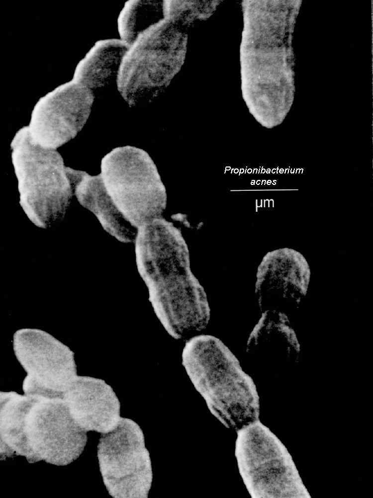
Case 1. A 43/M with CML s/p stem cell transplantation 2 years ago who p/w a 1.5 year h/o chronic lymphocytic meningitis associated with obstructive hydrocephalus secondary to chronic CNS Propionebacterium acne infection

 
 Taken from: http://luskiewnik.strefa.pl/acne/propionibacterium_acnes_files/propioni.jpeg

1. Propionebacterium acne has been shown in case reports and case series to cause chronic meningitis even in patients with no history of neurosurgical procedure (read here). It is seen in both immunocompromised and immunocompetent individuals. In most cases, a mononuclear-predominant pleocytosis is appreciated from the CSF.

2. Another learning point is that, the diagnosis of Propionebacterium acne chronic meningitis in this case was aided by the use of next generation sequencing (which maybe more sensitive that 16S rRNA gene sequencing). Read here regarding the NEJM article that chronicles how a patient with chronic neuroleptospirosis was diagnosed by this method.

Case 2. A 15-month old child with cartilage-hair hypoplasia syndrome (associated with severe combined immunodeficiency) presents with persistent diarrhea secondary to Norovirus infection

 Taken from: http://upload.wikimedia.org/wikipedia/commons/a/ae/Norovirus_4.jpg

1. Norovirus infection is self-limited and typically lasts for 2-3 days. However, in immunocompromised patients, the diarrhea can persist for several weeks to months and is sometimes associated with substantial symptoms that include severe debilitation and weight loss. It should be added in the differential diagnoses of immunocompromised patients with chronic diarrhea that eludes diagnosis. Further readings here and here.

2. Norovirus is diagnosed with stool PCR. There is no specific treatment for infection apart from supportive care. In immunocompromised patients, reduction of immunosuppression may help. Good evidence with the use of enteral immunoglobulins, nitazoxanide, and ribavirin is lacking at present.

Case 3.  A 68/F with cochlear implants presents with Streptococcus pneumoniae meningitis, pneumonia, and endocarditis (the Austrian triad/syndrome)

1. One needs to rule out endocarditis with a TEE in a patient with co-occurrence of pneumococcal meningitis and pneumonia. At least 2/3 of patients with pneumococcal endocarditis have concurrent pneumococcal pneumonia and meningitis.

2. Patients who develop acute otitis media after cochlear implantation have the greatest risk of subsequent pneumococcal meningitis. Most common organisms isolated are Streptococcus pneumoniae, Haemophilus influenzae, Streptococcus pyogenes, Branhamella catarrhalis, Staphylococcus aureus, mixed organisms, and gram negative bacilli.

3. Patients with cochlear implants and immunocompromising conditions (and recently, patients at least 65 years, read here) should get 13-valent pneumococcal conjugate vaccine on top of 23-valent pneumococcal polysaccharide vaccine.