Tuesday, November 4, 2014

November 4, 2014 Grand Rounds

Case # 1. A 55 year old white male who p/w a 2 month h/o headache, progressive mental status deterioration, persistent pleocytosis and evidence of leptomeningeal enhancement on MRI secondary to tuberculous meningitis.

Taken from:  http://www.bioquellus.com/interface/assets/images/content/Mycobacterium_tuberculosis_14313982_1.jpg

1. The case was diagnosed late in the course of the disease as all infectious disease work-up had been unremarkable. A prior brain and meningeal biopsy were also non-revealing. The diagnosis was only made when a second meningeal and brain biopsy revealed sparse evidence of AFB associated with granulomatous changes. Despite initiation of anti-TB treatment, he expired.

2. The case illustrates how hard it is to suspect and diagnose tuberculosis among persons living in low endemic areas. CNS tuberculosis carries a high mortality rate. Delay in treatment (even for a few days) is associated with mortality. If a high index of suspicion exists, treatment should be initiated while waiting for work-up.

3. To increase the sensitivity of CSF AFB analysis, a minimum of 3 serial lumbar punctures should be performed. This has been reported to increase CSF AFB sensitivity from 37 to 87% (read here).

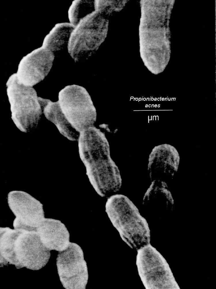
4. This case also highlights the potential use of next-generation sequencing to detect occult infection as illustrated by our Propionebacterium acne chronic meningitis case from Oct 14, 2014 and by the NEJM case of neuroleptospirosis as previously referenced.

5. Treatment of CNS tuberculosis (and pericarditis), in most cases, requires use of adjunctive corticosteroids to prevent immune reconstitution syndrome (so called 'paradoxical reaction') which can be life-threatening.

Case # 2. A 2 month old infant p/w fever, respiratory failure, hepatosplenomegaly, bilateral hydronephrosis, urinary infection from Serratia, and pneumonia from Burkholderia secondary to chronic granulomatous disease (CGD).

Taken from: http://www.immunopaedia.org.za/fileadmin/gallery/Chronic%20Granulomatous%20Disease/chronicgranulomatousdisease1.jpg 

1. CGD is caused by a genetic defect that prevents generation of supreroxide radicals that fight infection.

2. Remember that the autosomal recessive variant of CGD can occur later in life (as adolescents or adults).

3. Suspect CGD in an apparently healthy patient with recurrent or severe infection with Serratia, Burkholderia, Nocardia, Aspergillus, Salmonella, and Bacillus Calmette-Guerin (BCG).

4. One peculiar feature of CGD is the formation of granulomas that lead to visceral obstruction (e.g. small bowel obstruction, hydroureter, hydronephrosis).

5. CGD can be associated with the occurrence of  hemophagocytic lymphohistiocytosis (HLH) and autoimmune conditions. CGD can mimic systemic lupus erythematosus (SLE) and inflammatory bowel disease.

6. The use of Bactrim, itraconazole, and interferon gamma is the cornerstone of prophylaxis against infections among persons with CGD.

Tuesday, October 28, 2014

October 28, 2014 Grand Rounds

Case # 1. A 46/M with Burkitt's lymphoma, treatment-related myelodysplastic syndrome, s/p allogeneic stem cell transplantation 11 months prior to admission, who p/w intractable complex partial seizure and fever secondary to HHV-6 encephalitis

https://www.google.com/search?q=HHV-6&source=lnms&tbm=isch&sa=X&ei=natPVLyEMouvyQSZnYEo&ved=0CAkQ_AUoAg&biw=1920&bih=943#facrc=_&imgdii=_&imgrc=FqMPqQ62M74lAM%253A%3B9mqEToEvcY-DhM%3Bhttp%253A%252F%252Fupload.wikimedia.org%252Fwikipedia%252Fcommons%252Fc%252Fc8%252FHHV-6_inclusion_bodies.jpg%3Bhttp%253A%252F%252Fen.wikipedia.org%252Fwiki%252FHuman_herpesvirus_6%3B1350%3B900

1. The most consistently reported syndrome associated with HHV-6 infection among transplant patients is encephalitis. Patients can have mental status changes, behavioral disturbance, memory loss, and seizures. Other manifestations of HHV-6 in this patient population include: fever and rash, hepatitis, gastro-duodenitis, colitis, pneumonitis, and encephalitis.

2. A positive HHV-6 PCR from tissues can suggest the presence of disease. However, there is no single test that can differentiate HHV-6 causing disease from HHV-6 that is genomically integrated. A persistently high titer of HHV-6 PCR from tissues suggests genomically integrated HHV-6.

3. One study mentioned that even in asymptomatic transplant patients, HHV-6 can be detected in the CSF.

4. Treatment involves use of either ganciclovir, foscarnet, or cidofovir. There are no randomized controlled trials on antiviral efficacy. Reduction of a patient's immunosuppression is also recommended.Read here for more information.

Case # 2. A 31-day old infant presents with fever, rash, seizure, abdominal distension, and respiratory failure secondary to human parechovirus (HPeV) infection.

1. In an infant who presents with sepsis and CNS symptoms and the CSF is normal, think about HPeV infection. In one case series, CSF abnormalities in patients with HPeV infection were rare.

2. In adults, HPeV can present as fever, myalgia, and pharyngitis.

3. More on HPeV infection from blog entry dated July 22, 2014.

Tuesday, October 14, 2014

October 14, 2014 Grand Rounds

Case 1. A 43/M with CML s/p stem cell transplantation 2 years ago who p/w a 1.5 year h/o chronic lymphocytic meningitis associated with obstructive hydrocephalus secondary to chronic CNS Propionebacterium acne infection

 
 Taken from: http://luskiewnik.strefa.pl/acne/propionibacterium_acnes_files/propioni.jpeg

1. Propionebacterium acne has been shown in case reports and case series to cause chronic meningitis even in patients with no history of neurosurgical procedure (read here). It is seen in both immunocompromised and immunocompetent individuals. In most cases, a mononuclear-predominant pleocytosis is appreciated from the CSF.

2. Another learning point is that, the diagnosis of Propionebacterium acne chronic meningitis in this case was aided by the use of next generation sequencing (which maybe more sensitive that 16S rRNA gene sequencing). Read here regarding the NEJM article that chronicles how a patient with chronic neuroleptospirosis was diagnosed by this method.

Case 2. A 15-month old child with cartilage-hair hypoplasia syndrome (associated with severe combined immunodeficiency) presents with persistent diarrhea secondary to Norovirus infection

 Taken from: http://upload.wikimedia.org/wikipedia/commons/a/ae/Norovirus_4.jpg

1. Norovirus infection is self-limited and typically lasts for 2-3 days. However, in immunocompromised patients, the diarrhea can persist for several weeks to months and is sometimes associated with substantial symptoms that include severe debilitation and weight loss. It should be added in the differential diagnoses of immunocompromised patients with chronic diarrhea that eludes diagnosis. Further readings here and here.

2. Norovirus is diagnosed with stool PCR. There is no specific treatment for infection apart from supportive care. In immunocompromised patients, reduction of immunosuppression may help. Good evidence with the use of enteral immunoglobulins, nitazoxanide, and ribavirin is lacking at present.

Case 3.  A 68/F with cochlear implants presents with Streptococcus pneumoniae meningitis, pneumonia, and endocarditis (the Austrian triad/syndrome)

1. One needs to rule out endocarditis with a TEE in a patient with co-occurrence of pneumococcal meningitis and pneumonia. At least 2/3 of patients with pneumococcal endocarditis have concurrent pneumococcal pneumonia and meningitis.

2. Patients who develop acute otitis media after cochlear implantation have the greatest risk of subsequent pneumococcal meningitis. Most common organisms isolated are Streptococcus pneumoniae, Haemophilus influenzae, Streptococcus pyogenes, Branhamella catarrhalis, Staphylococcus aureus, mixed organisms, and gram negative bacilli.

3. Patients with cochlear implants and immunocompromising conditions (and recently, patients at least 65 years, read here) should get 13-valent pneumococcal conjugate vaccine on top of 23-valent pneumococcal polysaccharide vaccine.

Tuesday, September 16, 2014

September 16, 2014 Grand Rounds

Case # 1. A 54/M with chronic malaise, low back pain and new-onset heart failure secondary to subacute infected endocarditis (IE) secondary to Abiotrophia defectiva.

Taken from: http://medicine.creighton.edu/medschool/WebAtlas/secure/id/strep1/images/Strep%20NDS.jpg

1. Abiotrophia defectiva and Granulicatella sp are nutritionally-variant Streptococcus (NVS) that require pyridoxal for growth on culture. They are part of the normal oral, gastrointestinal, upper respiratory and urogenital flora. They are known to cause IE but they can also lead to primary bacteremia in patients with hematologic malignancy.

2. Compared with IE caused by viridans group Streptococcus, IE associated with NVS is usually characterized by smaller vegetation but with greater incidence of septic embolization. It also carries higher failure, relapse, and mortality rates. 

3. IE associated with NVS is treated with ampicillin and gentamicin (or vancomycin plus gentamicin for penicillin-allergic patients) similar to Enterococcus-associated IE. 

4. Abiotrophia defectiva shows in vitro susceptibility to vancomycin, rifampin, clindamycin, chloramphenicol, erythromycin, and levofloxacin. On the other hand, Granulicatella sp. are more likely to be resistant to penicillin and cephalosporins compared with Abiotrophia.


Case # 2. An 18/M with h/o bilateral lung transplantation for idiopathic bronchiolitis obliterans 2 years ago and poorly adherent to prednisone, tacrolimus, mycophenolate, and atovaquone presented with acute and progressive respiratory failure secondary to acute rejection; respiratory multiplex PCR tested positive for rhinovirus/enterovirus.

1. From last week's discussion, we know that the detection of rhinovirus/enterovirus using the multiplex PCR test is a clue to the presence of enterovirus D68. Because the patient had respiratory failure, further sequencing was done on the isolate. The isolate was identified as human rhinovirus 36.

2. Rhinovirus, unlike enterovirus, is tropic to the upper and not the lower respiratory tract. However, rarely, it can cause lower respiratory tract disease. Infection with rhinovirus is not associated with acute lung rejection in one case series.


Case # 3. A 30 year old man with well controlled HIV infection on HAART presented with acute rash that involved his bilateral palms and soles as well as acute Bell's palsy. 

Taken from: http://upload.wikimedia.org/wikipedia/commons/2/29/Treponema_pallidum.jpg

1. Bell's palsy occurs in secondary and tertiary forms of syphilis hence, performing a lumbar puncture to rule out neurosyphilis is warranted.

2. This patient had a normal head CT scan and lumbar puncture revealed 0 white cells and normal total protein. The absence of white cells in the CSF can rule out neurosyphilis but a more definitive test (although not standardized) that rules this out is a negative CSF FTA. The latter is usually ordered when the clinical suspicion for neurosyphilis remains in the presence of mild CSF pleocytosis and a negative CSF VDRL.

3. Read more about other unusual manifestations of secondary syphilis (i.e. gastropathy, hepatitis, Bell's palsy, aseptic meningitis ) here.

Tuesday, September 9, 2014

September 9, 2014 Grand Rounds

Case #1. A 70 year old man with relapsed AML undergoing chemotherapy admitted with fever, fatigue, sore throat, and cough; CT scan showed tracheal enhancement and mediastinitis; bronchoscopy showed pseudomembranous lesions; a case of invasive trancheobronchial aspergillosis

 Taken from: http://www.aspergillus.org.uk/secure/image_library/tracheobronch/fig7.jpg

1. Remember the different manifestations of aspergillosis in immunocompromised patients: pulmonary (from acute nodular lung disease to the more indolent necrotizing/cavitary form), tracheobronchitis, disseminated form, and rhinosinusitis. Less common manifestations that are well described in the literature include central nervous system involvement (brain abscess), endophthalmitis, endocarditis, gastrointestinal, or cutaneous lesions.

2. Invasive tracheobronchial aspergillosis (ITBA) is seen in <10% of patients with invasive aspergillosis. It is usually diagnosed late  because it can have normal imaging early in its course. A predisposed patient who comes in with fever, significant cough, and shortness of breath without pneumonia on chest CT scan should alert you to this diagnosis. Bronchoscopy is the best way to look for it.

3. ITBA has 3 forms: ulcerative (seen in AIDS and heart-lung transplant patients), pseudomembranous (seen in patients with hematologic malignancies), and obstructive (mucus plugs are usually seen but no bronchial inflammation is evident). Mixed forms can also be observed in <10% of patients with ITBA.

4.    ITBA is usually associated with a negative blood galactomannan.


Case # 2. A 15 year old girl presented with acute onset back pain and fever secondary to Group A Streptococcus epidural abscess

 http://images.ddccdn.com/images/pills/mtm/Clindamycin%20300%20mg-RAN.jpg

1. Always keep epidural abscess in your differential diagnosis for fever and back pain especially if patients have neurologic deficits. Be aware of the different stages of epidural abscess symptomatology: stage I (back pain, fever, tenderness), stage II (spinal root signs such as radicular pain), stage III (sensory and motor deficits, bladder/bowel dysfunction), and stage IV (paralysis).

2. This patient was treated with both ceftriaxone and clindamycin initially. The basis of combining clindamycin with penicillin in the treatment of Streptococcus pyogenes infection (especially in cases of high burden infection) early in its course is not only to prevent toxin release (in cases of necrotizing fasciitis) but also to counteract the theoretical Eagle effect. The latter is the paradoxical reduction of the bactericidal activity of high dose penicillin in infections with heavy Streptococcus pyogenes burden. The proposed mechanism is the reduced expression of penicillin-binding proteins by bacteria induced to a stationary phase of growth by the heavy bacterial burden. Clindamycin kills these bacteria in the stationary phase as its ability to inhibit bacterial protein synthesis is not dependent on bacterial growth stage or bacterial load.


Case # 3. Update on enterovirus D68

1. Enterovirus D68 was first isolated in 1962 in California. Since then, sporadic outbreaks have been seen. From 2008-2010, outbreaks have been documented in the Netherlands, Japan, China, and the Philippines. It has rarely been reported in the US until last month (read here).

2. Enterovirus D68 manifests almost exclusively as a respiratory illness of varying severity (from mild illness to frank respiratory failure requiring ventilatory support or extacorporeal membrane oxygenation). Rare cases have been associated with neurologic manifestations (e.g. flaccid paralysis and meningoencephalitis).

3. The clinician should be alerted of this entity when a multiplex PCR give out a result "enterovirus/rhinovirus". The virology lab can be called for genotypic identification as this is the only way to diagnose enterovirus D68. 

4. There are no specific treatments available for enterovirus D68 infection.

Tuesday, September 2, 2014

September 2, 2014 Grand Rounds

Case # 1 A 47 year old immunocompetent man with no significant past medical history who presented with  6 weeks of headache, neck pain, and fever secondary to Cryptococcus neoformans infection

1. In immunocompetent patients, Cryptococcus neoformans usually affects the central nervous system causing subacute to chronic meningitis. Patients often present with headache that is repeatedly misdiagnosed as migraine or cluster headache. 

2. Apart from HIV infection and receipt of immunocompromising medications (for transplantation, cancer, or rheumatologic disorders), other well-characterized immunodeficiency states that predispose to cryptococcal infection include CD4 lymphopenia and presence of anti-GM-CSF autoantibodies (read here).

3. Further ID pearls about cryptococcal infection are listed under Case # 1 June 24, 2014.


Case # 2. A 7-day old baby girl who presented with increased sleepiness, poor feeding, thrombocytopenia, and a diffuse macular rash secondary to parvovirus B19 infection


1. Parvovirus B19 causes fifth disease (erythema infectiosum). Another well-characterized but rarer and hence, less recognized illness that is associated with a rash secondary to parvovirus B19 is called papular-purpuric gloves and socks syndrome (PPGSS). PPGSS (see picture) is characterized by symmetrical and well-demarcated erythematous lesions on the hands and feet that end abruptly at the level of the wrist and ankle. It can present with high fever, mucosal lesions, and arthralgia (read here).

2. Other well-known illnesses associated with parvovirus B19 include: symmetric arthritis/arthralgia, transient aplastic crisis, chronic pure red cell aplasia, and non-immune hydrops fetalis. Rare manifestations include: acute hepatitis, fulminant liver failure, immune-complex glomerulonephritis, myocarditis, ITP, and TTP.


Case # 3. A 30 year old woman who is 13 weeks pregnant and who has a h/o splenectomy secondary to hereditary spherocytosis presented with fever and acute-onset watery diarrhea 2 days after eating in a restaurant secondary to Campylobacter jejuni infection (blood and stool culture positive)

1. Campylobacter usually causes a self-limited gastroenteritis and hence, treatment is not usually advised. However, treatment is recommended for those who present with severe disease (i.e. bloody diarrhea, high fever, extraintestinal infection, worsening or relapsing symptoms, or symptoms > 1 week duration) or those at risk for severe disease (e.g. immunocompromised, elderly, and pregnant patients).

2. The prevalence of fluoroquinolone-resistant Campylobacter is rising. In Thailand and Spain, for example, the prevalence is as high as 80%. In the US, fluoroquinolone-resistant Campylobacter has also risen from 0% to 19% between 1989 and 2001. Hence, some authorities advocate a macrolide (azithromycin or erythromycin) as first line agent against this infection.

3. Other antibiotics that can be used against Campylobacter include: aminoglycosides, carbapenem (especially for severely ill patients), clindamycin, tetracyclines, and chloramphenicol. It is inherently resistant to trimethoprim and some beta-lactam antibiotics including penicillin and most cephalosporins.

4. Campylobacter fetus is an uncommon species of Campylobacter that usually affects immunocompromised hosts, including elderly and pregnant women, and causes bacteremia and meningitis.

5. More ID pearls about Campylobacter are listed under Case # 2 July 2, 2014.

Tuesday, August 26, 2014

ID Grand Rounds. August 26, 2014

Case # 1. A 65 year old, CMV D+/R- kidney transplant recipient receiving mycophenolate and prednisone presented with 1 month h/o hematuria and progressive renal failure secondary to adenovirus nephritis.

From: https://www.google.com/search?q=adenovirus+electron+microscopy&source=lnms&tbm=isch&sa=X&ei=RP_8U9GlGsO0yAS5lICQCQ&ved=0CAgQ_AUoAQ&biw=1920&bih=943#facrc=_&imgdii=_&imgrc=jysh9OgcvqH5YM%253A%3BqFxYXFIK5hJFGM%3Bhttp%253A%252F%252Fwww.virology.net%252Fbig_virology%252FEM%252FAdeno-FD.jpg%3Bhttp%253A%252F%252Fwww.virology.net%252Fbig_virology%252FBVDNAadeno.html%3B979%3B964

1. Know the different clinical manifestations of adenovirus infection in immunocompromised patients: pneumonia (including necrotizing pneumonia and diffuse alveolar damage), gastroenteritis, hepatitis, hemorrhagic cystitis, interstitial nephritis, and meningoencephalitis. Among immunocompetent patients, adenovirus infection usually manifests as nonspecific upper respiratory tract illness, gastroenteritis, pharyngo-conjunctival fever, and epidemic keratoconjunctivitis.

2. Kidney transplant recipients who developed adenovirus graft-nephritis had better survival compared to other transplant recipients who developed native-kidney adenovirus nephritis according to one case series.

3. Histopathologic examination of a biopsy specimen is the gold standard for diagnosing invasive adenoviral infection. In situ hybridization is utilized to confirm the presence of the virus in tissues. Cells infected with adenovirus have large nuclei with basophilic inclusions and a thin rim of cytoplasm (so called smudge cells). Granulomatous formation is often observed. Adenovirus PCR can also be used but it should always be correlated with histopathology and with the clinical presentation to distinguish between asymptomatic infection and disease.

4. In immunosuppressed patients, the cornerstone of management for invasive adenovirus infection is supportive care and reduction of immunosuppression (read here). If treatment is desired, cidofovir is likely the best antiviral agent to use. Probenecid is used together with cidofovir to block active renal tubular secretion of cidofovir, thereby, preventing the development of nephrotoxicity. Aggressive hydration with fluid is recommended with cidofovir use.

5. In patients who develop adenovirus nephritis, concurrent use of probenecid is not recommended to ensure increased drug delivery to the site of infection.


Case # 2. A 6 year old child presented with 1 day of fever, diffuse maculopapular rash, and knee pain after returning from a 1 month trip to Puerto Rico a few days prior to admission secondary to chikungunya fever.

From: http://www.cdc.gov/chikungunya/images/maps/CHIK_Americas_Map-081214.jpg

1. Chikungunya ("that which bends up" in Tanzanian), was first described from an outbreak of fever in Tanzania in 1952.

2. It is characterized by fever that lasts for 3-5 days followed 2-5 days later by polyarthralgia (hands > wrists > ankles) that can sometimes be extremely debilitating. A diffuse maculopapular rash (which is pruritic in up to 50%) occurs in 40-75% of patients.  After the acute illness, up to 60% of patients can have persistent joint pains for up to 36 months.

3. The closest differential diagnosis is dengue fever as it is also transmitted by the Aedes mosquito vector. The main difference between dengue and chikungunya fever is that myalgia and polyarthralgia are virtually present in all chikungunya patients while they are uncommon findings in dengue patients. Thrombocytopenia is also more severe in dengue fever.

4. If the illness is suspected early (<5 days), obtaining plasma for viral PCR testing is the best method to diagnose chikungunya fever. Plasma viral-specific IgM and neutralizing antibodies are usually checked after 5 days of illness. Samples should be sent to the state laboratory or the CDC.

5. Outbreaks have traditionally been localized in Africa, Asia, and Europe but in December 2013, for the first time, chikungunya virus was found in the Americas. At present, there is an outbreak of chikungunya fever in the Caribbean. In July 2014, 2 patients from Florida who had not traveled outside of the US were diagnosed with chikungunya fever as reported by the CDC (read here).


Case # 3. A 21 year old woman from India develops fever, myalgia, diarrhea, severe leucopenia/thrombocytopenia, rhabdomyolysis, hepatitis, and multi-organ failure secondary to ciprofloxacin-resistant Salmonella enterica serotype Typhi infection (typhoid fever)

1. Tyhpoid fever can cause bone marrow suppression either by direct infiltration or induction of the macrophage activating system (hemophagocytic syndrome). Salmonella in the bone marrow can also lead to granuloma formation.

2. Alternative antibiotics used for the treatment of ciprofloxacin-resistant Salmonella include: ceftriaxone, azithromycin, or chloramphenicol. Other agents that may be active include imipenem and trimethoprim-sulfamethoxazole.

Tuesday, August 19, 2014

August 19, 2014 Grand Rounds

Case # 1. A 35/F presented with 4 month history of fever, diarrhea, vomiting, 30 pound weight loss, and granulomatous ileitis 

1. Several differential diagnoses were brought up including tuberculosis, histoplasmosis, Crohn's disease, sarcoidosis, and lymphoma. A useful article to review the different causes of granulomatous disease is linked to Case # 3 from June, 17, 2014.

2. It is hard to distinguish Crohn's disease from gastrointestinal tuberculosis especially in areas where the prevalence of both diseases is high. A positive anti-Saccharomyces cerevesiae (ASCA) antibody test is common in patients with Crohn's disease while a positive tuberculin skin test (TST) or interferon gamma release assay can be supportive of tuberculosis. Tissue culture remains the gold standard of diagnosing gastrointestinal tuberculosis.

3. Again, it was emphasized (similar to Case # 3 from June 17, 2014) that multiple tissue biopsies and cultures should be obtained to determine the cause of the granulomatous process. Empiric anti-tuberculous treatment may be warranted in some patients if the index of suspicion for tuberculosis is high.


Case # 2. A 7-week old infant presented with 1 day history of irritability, low grade fever, tachycardia, and localized neck cellulitis and adenitis secondary to group B Streptococcus (GBS)

From: http://www.giglig.com/wp-content/uploads/2011/09/Streptococcus.jpg

1. GBS cellulitis-adenitis syndrome is a rare (34 cases in the literature) but well-described late-onset GBS disease in neonates and infants. The median age of onset is 4.5 weeks. It usually presents as fever, increased irritability, and poor feeding followed by the appearance of lymphadenitis that is usually located in the submandibular region. In >90% of cases, GBS is isolated from the blood or from the lymph node.  


Case # 3. A 58/F presented with acute onset fever, nausea, vomiting, diarrhea, leukocytosis to 44,000, enterocolitis on CT scan, and septic shock several weeks after completing levofloxacin treatment for community acquired pneumonia; stool C. difficile toxin test is negative on two occasions

From: https://ndnr.com/wp-content/uploads/2012/01/10045000_m.jpg

1. The sensitivity of Clostridium difficile toxin enzyme immunoassay (EIA) is 63-94% depending on the the commercial test used (read here). The EIA we use for detecting Clostridium difficile toxin in our hospital is called TECHLAB assay. From the package insert, this test, compared to tissue culture cytotoxicity assay which is the gold standard in diagnosing Clostridium difficile, has a sensitivity of 92.2%, specificity of 100%, negative predictive value of 98.6%, and positive predictive value of 100% (read here).

2. The EIA can have as much as 10-20% false negative rate (read here). Thus, in some rare cases when this test is negative, it may be reasonable to continue treatment for Clostridium difficile. Although, in these cases, one should always look for other competing diagnoses such as ischemic bowel disease and other antibiotic-associated enterocolitis (e.g. Campylobacter, Salmonella).

Wednesday, August 13, 2014

August 12, 2014 Grand Rounds

Case # 1. A 19 year old African American man who presents with a recent history of proven HSV encephalitis treated with 21 days of intravenous acyclovir presented with recurrent neurological symptoms and worsening brain MRI findings secondary to recurrent versus refractory HSV.

From: http://posterng.netkey.at/esr/viewing/index.php?module=viewimage&task=&mediafile_id=508165&201302022314.gif

1. In most HSV encephalitis cases, 21 days of acyclovir may be enough. However, in cases where the initial infection is severe, like in this patient with extensive edema of the temporal lobe and serious neurological manifestations, treating for more than 21 days may be beneficial. We can learn from how our Pediatric Infectious Disease group here treat neonatal HSV encephalitis. On the 19th day of acyclovir treatment, a repeat lumbar puncture is performed and HSV PCR is rechecked from the CSF. If this comes back positive for HSV, treatment is extended for another 1-2 weeks.

2. Relapses of HSV encephalitis are usually seen in the pediatric age group, especially in those patients treated with only 10 days of acyclovir.

3. Interestingly, another differential diagnosis for a patient with relapsing symptoms after treatment of HSV encephalitis is anti-NMDA receptor encephalitis! Infection with HSV in the brain triggers the production of NMDA receptor antibodies that are responsible for this syndrome. Read more here. This is not the case here as our patient has a positive CSF HSV PCR on readmission. 

4. Anti-NMDA receptor encephalitis is an autoimmune disorder usually seen among young women with an ovarian teratoma that is characterized by prominent psychiatric manifestations, autonomic instability, mutism, memory deficits, and dyskinesias (read here for a good review). Every infectious disease physician should know this diagnosis as its prevalence now surpasses that of viral encephalitis in the California Encephalitis Project cohort (read here). Anti-NMDA encephalitis is treated with steroids.

4. Recurrent HSV encephalitis merits a work-up for immunodeficiency. Toll-like receptor 3 (TLR3) deficiency and NK cell deficiency predispose patients to developing recurrent HSV encephalitis.


Case # 2. A 20 month old child presented with a 3-day history of an ulcerating lesion in the nose secondary to ecthyma gangrenosum as a result of Pseudomonas aeruginosa infection.

From: http://www.langetextbooks.com/0071774343/gallery/035_ch18.jpg

1. Ecthyma gangrenosum is not exclusive to Pseudomonas. Staphylococcus aureus, other gram negative bacteria, and fungi are also known to cause it. 

2. Ecthyma gangrenosusm starts out as a painless macule. It then rapidly develops induration and becomes pustular (sometimes bullous). Later on, it ulcerates and becomes gangrenous (with a central black eschar). The final appearance (as shown in the picture) is a lesion with a necrotic center surrounded by a usually raised red or violaceous border. The evolution from a macule to a necrotic ulcer typically takes 12-18 hours!


Case # 3. A 68 year old man with pure red cell aplasia of undetermined cause, history of recent alemtuzumab treatment, who presented with fever, chest pain, and pulmonary infiltrates associated with a pleural effusion secondary to Actinomyces naeslundii.

1. Alemtuzumab is an anti-CD52 monoclonal antibody used in many refractory lymphoproliferative diseases and autoimmune conditions. It causes a profound depletion of T cells that lasts for many months. Its use has been associated with development of a variety of opportunistic (especially CMV, PML, adenovirus, toxoplasmosis) and non-opportunistic infections (e.g. bloodstream infections). Read more here.

2. We often think of actinomycosis when the infection is said to spread beyond tissue planes. Well, if caught early, like in this case, we can just sometimes appreciate localized disease (in this case, empyema without chest wall extension).

3. Remember that even if Actinomyces is an anaerobic bacteria, it is resistant to metronidazole.

Wednesday, August 6, 2014

August 5, 2014 Grand Rounds

Case 1. 52 year old woman with SLE who presents with chronic progressive headache, diplopia, blurring of vision, numbness of her toes, and constitutional symptoms secondary to histoplasmosis.

 From: https://blogger.googleusercontent.com/img/b/R29vZ2xl/AVvXsEixsT5aJ5G8uBhS4qVZHCyssDojsaqfcvKBj0wnpJ7fwG6WzN6wuFxqrfG1dsTS3zD9rcbGz4wD4dA2aDZEdJcTPMeto4ifEHU-xZyT5-cLe86qSxO1dmSUfnBan6AIxh93JQ35ebEtkkw/s1600/fungal+distribution.png

1. Urine Histoplasma antigen cross-reacts with blastomycosis, paracoccidioidomycosis, penicilliosis, coccidioidomycosis, and African histoplasmosis. Only blastomycosis is endemic in Missouri. Treatment of histoplasmosis is very similar to that of blastomycosis.

2. Blastomyces antibody testing, which is usually done by an immunodiffusion assay, has low sensitivity but relatively good specificity. It cross-reacts with histoplasmosis, however. 

3. Distinguishing blastomycosis and histoplasmosis may be difficult and sometimes rely on isolation of the actual fungus from a tissue specimen. Clinically, they can present similarly as well. However, there are some important distinctions.
  • Pulmonary findings are identical (from solitary to multiple lesions, from nodular to cavitary infiltrates).
  • Skin findings may help differentiate the two. Histoplasma can give rise to almost any skin findings. Erythema nodosum and erythema multiforme are common. Blastomyces, on the other hand, presents usually with a short list of skin manifestations. This includes pyogranulomatous lesions with pseudoepitheliomatous hyperplasia (sometimes misdiagnosed as skin cancer). It can also present with ulcerating or verrucous lesions or cold abscesses. 
  • Brain abscess and bone and joint infection are also more prominent clinical manifestations of blastomycosis.
  • To confuse you further, African histoplasmosis (Histoplasma capsulatum var duboisii) presents exactly like blastomycosis (in terms of bulky skin lesions and prominent bone and joint involvement).

Case 2. A 17 year old girl who presents with acute exudative tonsillopharyngitis, fever, and pneumonia secondary to Lemierre's syndrome caused by Fusobacterium necrophorum.

From: http://iam.uic.edu/wp-content/uploads/2012/03/Lemierre-Syndrome-2.jpg

1. Sore throat followed by pneumonia, especially where chest imaging suggests possible septic emolization, should raise suspicion for Lemierre's syndrome. In this situation, get a Doppler ultrasound of the neck to look for internal jugular vein thrombosis.

2. Anticoagulation in cases of Lemierre's syndrome is still a controversial topic. At least two case series of Lemierre's syndrome showed no difference in outcome among patients who received anticoagulation compared to those who did not.

Case 3. A 73 year old man with history of recurrent and metastatic cholangiocarcinoma presents with multiple metastatic versus septic embolization of the liver, spleen, and kidneys as well as a new 3+ aortic regurgitation associated with an aortic valve vegetation.

From: http://classconnection.s3.amazonaws.com/695/flashcards/739695/jpg/me1316628051717.jpg

1. Nonbacterial thrombotic endocarditis (NBTE) (formerly known as marantic endocarditis) are most commonly seen in patients with advanced malignancy (usually lung and gastrointestinal cancer). It can also be seen in patients with rheumatologic conditions. 

2. In a case series of cancer patients with NBTE, one-third had mild to moderate valvular regurgitation (look here).

3. It is very difficult to distinguish NBTE from culture-negative endocarditis. Obtaining a specimen of the valve and the vegetation, which is an impractical approach especially for cancer patients who are not good candidates for surgery, may sometimes be the only way to differentiate the two.

Tuesday, July 29, 2014

July 29, 2014 Grand Rounds

Case # 1. A 19 year old man with diffuse macular rash, bilateral red eyes, fever, headache, abdominal pain, diarrhea, elevated liver enzymes and leukocytosis secondary to acute leptospirosis
 http://0101.nccdn.net/1_5/317/170/030/leptospira.jpg

1. Remember that not all patients with leptospirosis present with the deadliest form of the infection called Weil's syndrome (i.e. profound jaundice with total bilirubin out of proportion to the liver enzyme elevation, renal failure, pulmonary hemorrhage, hemorrhagic diathesis). Majority of patients present with the anicteric form of the disease.

2. There are usually two phases of anicteric leptospirosis (although, an overlap between the two commonly occurs):

     A. Septicemic phase.  Characterized by abrupt onset of fever, headache, chills, myalgia, conjunctival suffusion, nausea, vomiting, diarrhea, and rash. Lasts for 5-7 days.
  • Conjunctival suffusion. Manifests as bilateral conjunctivitis without eye discharge (eyes appear "wet"). Although not specific for leptospirosis, its presence in a patient with nonspecific symptoms should raise suspicion for leptospirosis. In fact, the World Health Organization (WHO) includes this in its scoring system for diagnosing leptospirosis (read here).
  • Myalgia is also a predominant symptom. Calf tenderness is most often cited in the literature and is also included in the WHO scoring system.
  • Rash occurs in 8-12% of patients. It is usually transient. Can appear as urticarial, macular, maculopapular, or purpuric.
     B. Immune phase. Characterized  by aseptic meningitis in 80% of patients. Uveitis, renal insufficiency, respiratory symptoms, and hepatosplenomegaly may occur.

3. The icteric form of leptospirosis (Weil's syndrome) is not usually biphasic. It usually manifests with persistent fever and abrupt onset of jaundice and profound renal failure.

4. The diagnosis rests on serologic testing. The CDC defines a positive serology as a titer of >/= 1:200 by the microscopic agglutination test (MAT). At Barnes-Jewish Hospital, the samples are sent to the Mayo Clinic which uses indirect hemagglutination assay (IHA) for diagnosing leptospirosis. For this test, the cut-off of a positive result is a titer of at least 1:50. A single titer of >/= 1:100 is suggestive of active or recent leptospirosis. The IHA is 100% sensitive and 97% specific for leptospirosis compared with the MAT.


Case # 2. A 3 year old girl coming from a trip to Cameroon, presents with fever, abdominal pain, vomiting, and diarrhea secondary to non-typhoidal Salmonella infection
 http://images.gizmag.com/hero/salmonella.png

1. Non-typhoidal Salmonella is the most common cause of bacteremia in Africa according to one study.

2. Non-typhoidal Salmonella infection is usually self-limited. Treatment is required for patients with severe symptoms (e.g high fever, prolonged diarrhea needing hospitalization), for immunocompromised patients (e.g. AIDS, organ transplant, use of steroids or other immunosuppressive agents, sickle cell or other hemoglobinopathies), patients > 50 years, and those with prosthesis or severe valvular heart disease.


Case # 3. A 24 year old man who presented with a 1 month history of diffuse erythrodermic scaly rash, fever, weight loss, night sweats, arthralgia, leukocytosis, and diffuse lymphadenopathy mistaken for an infectious process and Still's disease; an excision biopsy of an enlarged inguinal lymph node revealed classic Hodgkin lymphoma
 http://hematopathology.stanford.edu/images/HP-Home-Image.jpg

1. In a patient with symptoms consistent with lymphoma and unrevealing infectious disease work-up, always pursue excisional lymph node biopsy. Fine needle aspiration biopsy is inferior to excisional biopsy for diagnosing lymphoma. Lymphoma is a great mimic! There are many cases reported in the literature of occult lymphoma diagnosed only with several excisional lymph node biopsies.

Tuesday, July 22, 2014

ID GRAND ROUNDS: July 22, 2014

Case #1. A 20-day old girl with fever, irritation, elevated liver enzymes, and tachycardia secondary to human parechovirus infection.

Taken from http://jvi.asm.org/content/85/3/F1.medium.gif

1. Human parechovirus (HPeV) is genetically and phenotypically similar to enterovirus. Its clinical presentation is also indistinguishable from enterovirus. However, it is not detected by routine enterovirus PCR testing. In a child with enterovirus-like clinical disease but the enterovirus PCR is negative, suspect HPeV infection.

2. It commonly affects children < 2 years (only a few case reports of infection in children > 10 years). Most commonly manifests as meningoencephalitis, sepsis, hepatitis, and mycocarditis.

3. Treatment is supportive. Pleconaril does not have activity against HPeV. IVIG, which has been shown to be beneficial in some cases of enterovirus infection (e.g. myocarditis), has not been studied in HPeV infection. Pocapovir is a new agent against HPeV currently being studied.


Case # 2. A 64 year old woman from New Mexico, with end-stage liver disease, s/p orthotopic liver transplantation 2 months prior to admission, who presents with fever, chills, malaise, nausea, and vomiting; found to have pancytopenia secondary to Ehrlichia infection

Taken from  https://ahdc.vet.cornell.edu/sects/ClinPath/modules/CaseMonth/images/June2012/Figure%204.jpg

1. In a case series of 15 solid organ transplant recipients with ehrlichiosis, transplant patients experienced less rash and had lower liver enzymes compared to immunocompetent patients. However, they had more leucopenia and renal dysfunction. There were no deaths in this case series because doxycycline was administered within 48 hours of presentation. Read more on this here.


Case # 3. A 70 year old woman with leukemia, s/p stem cell transplantation 7 months prior to admission, who presents with refractory CMV disease and viremia secondary to multi-drug resistance


1. Ganciclovir, in order to be active against CMV, has to undergo several steps of phosphorylation by enzymes produced by CMV. Let's review CMV antiviral resistance.

    A. UL97 phosphotransferase mutation (inhibits initial step of ganciclovir phosphorylation); it is the most commonly seen CMV mutation; confers resistance to ganciclovir

    B. UL54 DNA polymerase mutation (inhibits succeeding steps of ganciclovir phosphorylation); it is less commonly seen compared to UL97 mutation; confers various combination of resistance to not only ganciclovir but also to foscarnet, and/or cidovir.

   C. The presence of both UL97 and UL54 mutations usually confers high-level resistance to ganciclovir. Isolated UL54 mutation (in the absence of UL97 mutation) is very uncommon.

2. It is important to monitor CMV viremia during treatment with ganciclovir. You should expect a twofold drop in viremia per day or at least a tenfold drop in 1 week. Significant log drop should be expected in 2 weeks. CMV viremia not responding to ganciclovir should be checked for UL97 and UL54 genotypic mutations.

3. Treatment options for drug-resistant CMV (adapted from Deepali Kumar's AST Handbook of Transplant Infections, 2011):

   A. Reduce immunosuppression. Sirolimus maybe used to replace other calcineurin inhibitors as it has been shown to have some degree of activity against CMV.

  B. CMV with only UL97 mutation. For low-level mutation, can use high dose ganciclovir. For high-level resistance, can use foscarnet alone or in combination with ganciclovir.

  C. CMV with UL54 mutation (again, this is usually associated with UL97 mutation). Can use foscarnet +/- CMV immunoglobulin. The other agent that can be used include CMX-001, leflunomide, artesunate, and maribavir.

4. Letermovir is a new drug against CMV infection (read here) currently being studied in phase II trials (for CMV prophylaxis, read here). It is a promising agent since it has a novel mechanism of action (inhibits CMV pUL56 gene thereby disrupting the viral terminase complex necessary for viral replication) and has low toxicity, good oral bioavailability, and low potential for drug-drug interactions.

Wednesday, July 16, 2014

July 15, 2014 Grand Rounds

Case 1: A 70 year old immunocompetent woman presenting with 4 weeks of generalized body weakness and gram positive bacilli that grew on 4 culture bottles on admission.

Taken from: http://www.easynotecards.com/uploads/977/95/69c3b13a_13a8788d737__8000_00000081.jpg

1. The gram positive bacilli in this case was later identified as Bacillus sp. (not anthracis). The same blood cultures also showed growth of methicillin-susceptible Staphylococcus aureus and Staphylococcus hominis.

2. Let's review the different clinically important gram positive bacilli (look here). Our laboratory routinely reports branching filamentous organisms (BFO). It is important to note that some gram positive bacilli are also AFB positive. As you can see from the diagram, the clinically significant gram positive bacilli are diverse and are responsible for a wide spectrum of illnesses.

3. Can Bacillus (not athracis) cause clinically significant infection? The answer is yes. Bacillus cereus is known to cause serious endophthalmitis. It has also been reported as a cause of endocarditis (see here, kuddos to our very own Ben Thomas). Bacillus bacteremia have been reported among intravenous drug users, preterm neonates, and those with long-term indwelling catheters.

Case 2: A 14 year old boy presenting with acute frontal head swelling and pain, rhinorrhea, cough, fever, and post-nasal discharge secondary to Streptococcus angionosus Pott's puffy tumor.

Taken from: http://www.asnr2.org/neurographics/8/3/68/What%27s%20in%20a%20Name_files/slide0029_image128.jpg

1. Pott's puffy tumor refers to frontal bone subperiosteal abscess associated with frontal bone osteomyelitis. It occurs as a complication of frontal sinusitis or trauma. Most patients with Pott's puffy tumor are immunocompetent. All patients present with frontal head swelling. Complications include intracranial extension (80%) and orbital cellulitis (30%).

2. Common etiologic agents for Pott's puffy tumor include Streptococcus (pneumoniae, angionosus, Group A), Staphylococcus (aureus usually), and anaerobes (Fusobacterium, Propionebacterium). Cultures can be polymicrobial or sterile as well.

3. Streptococcus angionosus group (once known as Streptococcus milleri) include Streptococcus angionosus, Streptococcus intermedius, and Streptococcus constellatus. They have a characteristic butterscotch smell on culture media. They are associated with abscess formation.

Case 3: This is a management case

A 22 year old man with paraplegia presenting with sepsis secondary to osteomyelitis and soft tissue infection from multi-drug resistant organisms (vancomycin-resistant Enterococcus; methicillin-susceptible Staphylococcus aureus; Pseudomonas aeruginosa susceptible only to colistin, amikacin, gentamicin, and tobramycin; and Candida lusitaniae)

1. The main point that was emphasized is that, management of chronic osteomyelitis in paraplegic patients is very challenging and one that requires a multidisciplinary approach. Antibiotics alone will not be sufficient.


Tuesday, July 8, 2014

July 8, 2014 Grand Rounds

Case # 1. A 24 year old man from Arkansas with no significant past medical history who presented with 9 days of fever, nausea, vomiting, diarrhea, abdominal pain, cholecystitis, and elevated liver enzymes secondary to acute histoplasmosis

Taken from http://wwwnc.cdc.gov/eid/article/17/9/10-1987-f1

1. Gastrointestinal (GI) histoplasmosis as a presenting symptoms of acute histoplasmosis is rare but is actually found in 70% of disseminated histoplasmosis on autopsy. It should be considered in the differential diagnosis of a patient with acute progressive gastroenteritis. The geographic distribution of histoplasmosis cases in the US is shown in the diagram.

2. Most common presentation of GI histoplasmosis include mucosal ulcers and abdominal lymphadenopathy. It can also present as ileitis that mimic inflammatory bowel disease (can add this in the differential diagnoses of infectious etiologies that cause ileitis such as tuberculosis, amoebiasis, Yersinia/Salmonella infection, and actinomycosis among others).

3. A negative Histoplasma antigen test does not rule out disseminated histoplasmosis. The urine Histoplasma antigen test is more informative in the immunocompromised host compared to the immunocompetent patient (read here).

4. Histoplasma antibody testing is more informative in immunocompetent hosts compared to immunocompromised patients where it can be falsely negative.

Case 2. A 2 week old preterm and small for gestational age infant with persistent methicillin-susceptible Staphylococcus aureus bacteremia

1. An aggressive search for any focus of infection is a priority in patients with persistent staphylococcal bacteremia. Have a low threshold for draining localized areas of fluid collections.

Case 3. A 59 year old woman with alcoholic cirrhosis, CMV colitis, recurrent profuse diarrhea,  respiratory failure, and pancytopenia who was found to have Pneumocycstis jirovecii pneumonia (PCP)

Taken from http://bestpractice.bmj.com/best-practice/images/bp/en-gb/19-4_default.jpg


1. Cirrhosis and severe malnutrition are enough to make one immunocompromised. Infection with opportunistic infections secondary to CMV and PCP have been reported in cirrhotic and malnourished patients. Include PCP in the differential diagnoses of refractory pneumonia even in non-AIDS and non-translant patients.

2. The presence of cystic lung disease and pneumatoceles, although not specific for any etiologic agent for pneumonia, is an  underrecognized radiolographic manifestation of PCP.

Tuesday, July 1, 2014

July 1, 2014 Grand Rounds

Case 1. 60 year old man with acute onset fever, confusion, renal failure, progressive pancytopenia, respiratory distress, and septic shock secondary to Heartland virus infection.

 (Picture taken from: http://upload.wikimedia.org/wikipedia/commons/2/29/Amblyomma_americanum_tick.jpg)

1. Heartland virus is a newly discovered phlebovirus (family Bunyaviridae) first described in 2009 in a farmer from northwestern Missouri who presented with fever, thrombocytopenia, and leucopenia. It is also transmitted by the Lone Star tick (Amblyomma americanum), the main vector of ehrlichiosis, tularemia, and Southern Tick Associated Rash Illness (STARI).

2. A patient who presents with fever, leucopenia, thrombocytopenia, and transaminitis but has repeatedly negative tests for Ehrlichia should be tested for Heartland virus infection (contact Dr. George Turabelidze at george.turabelidze@health.mo.gov of the Missouri Health Department). Heartland virus is diagnosed by a positive Heartland virus RNA or a fourfold rise of antibody titers between acute and convalescent samples.

3. The Heartland virus will not respond to doxycycline. No therapy is available.

4. Heartland virus infection can be lethal. Our case represents the 3rd mortality in the US (first mortality was from Tennessee, second mortality was from Oklahoma).

5. Heartland virus closely resembles the severe fever with thrombocytopenia syndrome virus recently described from China in 2007.

Case 2: A 15 year old male with acute abdominal pain, fever, and hypotension secondary to Campylobacter jejuni infection

1.This patient eventually developed diarrhea during his hospital stay. Patients with fever and diarrhea should always have a stool culture to rule out invasive bacterial infection secondary to Salmonella, Shigella, and Campylobacter. Other causes of fever and diarrhea include rotavirus, norovirus and Clostridium difficile infection.

2. Campylobacter infection can be associated with reactive arthritis (3% of cases), Guillain-Barre syndrome (<1% of cases), and bacteremia (<1% of cases). Reactive arthritis and Guillain-Barre syndrome usually manifest 1-2 weeks (occasionally several weeks) after onset of diarrhea.

Case 3. This is a management case. 

Patient with acute myelogenous leukemia who presents with multi-drug resistant Enterococcus infection (vancomycin resistant, susceptible only to linezolid and daptomycin), Candida glabrata (susceptible only to fluctosine, no susceptibility break point for amphotericin), and Stenotrophomonas maltophilia (susceptible only to minocycline).

1. Patient was treated with minocyline, amphotericin, and linezolid. Amphotericin-resistant Candida is exceedingly rare (except for Candida lusitaniae).

Tuesday, June 24, 2014

June 24, 2015 Grand Rounds

Case 1. This is a management dilemma case. 


A 45/M with h/o cryptococcal meningitis and recurrent admissions for headache relieved by lumbar puncture despite negative fungal cultures. The patient had more than 2 reinduction therapies with Ambisome followed by fluconazole consolidation/maintenance therapies. Despite this, the patient kept getting admitted for recurrent headache, CSF pleocytosis, mild to moderate CSF protein elevation, but negative fungal cultures.

1. Fluconazole-resistant Cryptococcus is rare. However in Africa, it is an emerging problem given that fluconazole prophylaxis and use of fluconazole monotherapy for cryptococcal meningitis is widespread. In a study done in South Africa (read here), two thirds of patients with symptomatic relapse had positive culture. Of these positive cultures, 76% had reduced fluconazole susceptibility.

2. Repeating cryptococcal antigen in the serum or CSF of this patient was suggested. The guideline (read here) explicitly mentioned that changing antigen titer (or positive India ink, or changes in CSF chemistries and cellular reactions) is insufficient in making a diagnosis of relapse. Let's go over some of the definitions:

      A. Relapse: Cryptococcus culture positive + recurrence of signs and symptoms; suggests prior
      clearance of cultures and resolution of signs symptoms;

      B. Persistence: persistence of positive Cryptococcus culture after 4 weeks of effective therapy at
      effective doses.

In this particular patient, measuring CSF cryptococcal antigen may be informative. Dr. Powderly and colleagues published in 1994 a study (read here) that showed that during therapy for acute meningitis, an unchanged (defined as 0-1 dilution change) or increased titer (defined as a rise of at least 2 dilutions) CSF antigen was correlated with clinical and microbiological failure especially among patients with baseline antigen titer of  > or = 1:8.

3. It was suggested that in the absence of good evidence of relapsed infection in this patient, chronic management of elevated intracranial pressure (e.g. VP shunt placement) may suffice without reinstituting induction therapy.

Case 2. 12 year old boy with common variable immunodeficiency (CVID) presenting with 3 months of fever, night sweats, and weight loss as well as multiple enhancing mediastinal and inguinal lymph nodes. Patient eventually found to have non-caseating granulomas associated with AFB+ organism on lymph node biopsy.


1. Tuberculosis and non-tuberculous Mycobateria infection are rare in patients with CVID. There is a short list of case reports, however.

2. Fifty percent of CVID patients develop complications: 20% of complicated cases have non-caseating granulomas, 50% have lymphoproliferative diseases, 20% have autoimmune diseases, and 4% have lymphoma.

3. Most common sites of granuloma formation are the lungs, spleen and liver. They tend to respond better with corticosteroid therapy compared to granulomas located elsewhere.

4. This patient was maintained on antibiotics that target both tuberculosis and non-tuberculous Mycobacteria since all his cultures were negative.

Case 3.This is another management dilemma case.


A 28/F with recurrent mitral valve lesion over 3 years. Patient had 3 different courses of infective endocarditis treatment and two major valve surgeries. Work-up for infection, autoimmune disease, and hypercoagulable state have been unrevelaing.

1. Let's review common causes of culture-negative endocarditis. In a case series of 348 patients with blood culture-negative endocarditis (read here), the following organisms were isolated by different testing methods: Coxiella (48% ), Bartonella (28%), Streptococci (4 cases), Tropheryma (2 cases), Abiotrophia (1 case), Mycoplasma (1 case), and Legionella (1 case).

2. The value of utilizing next generation sequencing in this patient was brought up, in reference to the case recently published in the New England Journal of Medicine (read here) and the New York Times (read here). The article tells the story of a 4-year old boy with recurrent meningitis and unrevealing tests. With the help of next generation sequencing, the boy was diagnosed with neuroleptospirosis.